
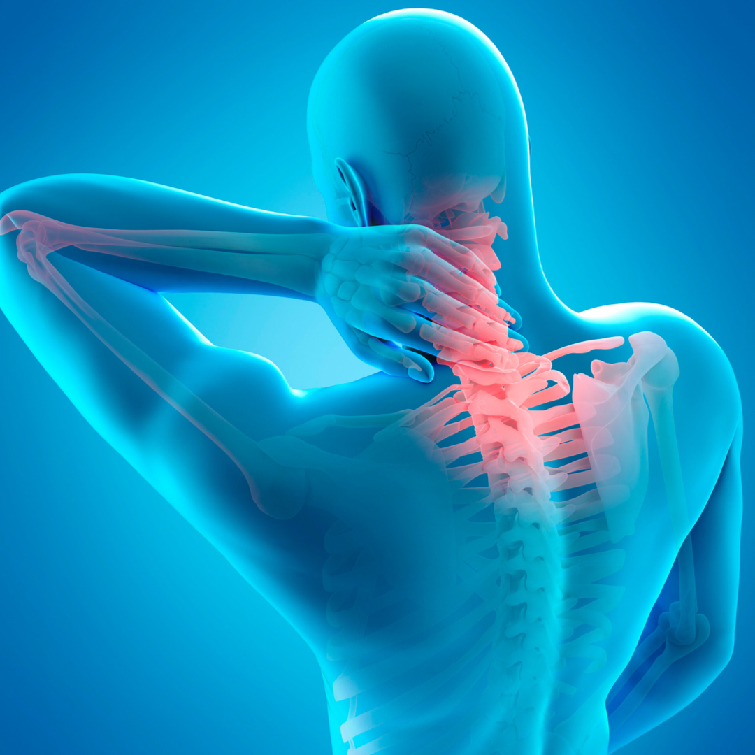
The cervical disc has been maligned, as far as cause and effect, in the last 20 years. The biggest culprit is the MRI, which creates a false level of diagnosis simply by diagnosing a degenerative disc, which is present at one or two levels in 85% of people over 45 or 50. Historically, a cervical disc was never diagnosed by an MRI. I practiced for 10 years before MRIs existed, and prior to that a “cervical disc” was diagnosed by radiculopathy, specific pressure on a nerve root causing pain down the arm. The C4-5, C5-6 or 6-7 are the most common levels. This was outlined by Robbie Robinson as a Chairman of Johns Hopkins back in the 1950s to explain what a cervical disc was, and 85% of the time, it was an osteophyte.
So cervical disc, as elucidated by Dr. Robinson in his landmark paper in 1955, delineating the Smith Robinson graft, a method of fusing the spine, was to eliminate the motion at that level, allowing the osteophyte to resolve, which occurred with excessive motion with the degeneration, and resolving the problem. So we are not treating neck pain, we’re treating arm pain. Arm pain is weakness of the deltoid muscle, which would be C4-5, weakness of the wrist, which would be C5-C6, weakness of the hands and grip or the triceps muscle which is C6-C7, which is caused by pressure on a nerve root. And this was classic. It would encompass pain or numbness or reflex changes or weakness of these muscle groups.
This is called radiculopathy. Radiculopathy is not a subjective complaint of hand numbness, which can be a myriad of other problems such as scalene syndrome. But back to the original discussion, lumbar disc surgery is done generally for a soft disc pushing up against a nerve root. That does not happen in the cervical spine, except in younger people on a rare instance. I, myself, have only seen a rare number in the last 40 years. As I said, it’s a degenerative phenomenon. A radiculopathy basically is how we diagnosed these things before MRIs existed. Pressure on a nerve root was determined clinically, not subjectively and not by image. And if it did not respond to usual conservative treatment; steroids, cervical traction; then a myelogram was done with a CT scan, which delineated where this bone spur or osteophyte was. The osteophyte will resolve, as per Dr. Robbie Robinson’s article in 1955 describing the Smith Robinson graft. Now, if degeneration occurs at that disc level and the MRI, which came out in 1988 or 1989 shows a degenerative disc, that doesn’t prove where the problem comes from. A disc itself, as it degenerates, starts to settle. As it settles is kind of like a car tire full of air, and the edges bulge out a little bit. This is read as “a bulging disc” by many non-astute radiologists, and enhances the decision-making for predatory surgeons.
A bulging disc can push up against the spinal cord. It can close the cerebral spinal fluid space, which is a small, thin white line on the MRI in front and in back of the spinal cord. Closure of this CSF space is irrelevant. Actually, pressure on the spinal cord can cause spinal cord changes, which can be asymptomatic and is debated as to whether that’s a significant problem. My criteria for that, as far as diagnostics, is a flexion-extension film at that level, a standing lateral flexion-extension film of the cervical spine. And if, let’s say, C5-C6 or C4-C5 does not move on a flexion-extension film and there’re no symptoms, then one would hold off on doing anything because it’s already fused. Whether there’re changes in the spinal cord, people might call that myelopathy, but without significant findings, it’s irrelevant.
What are significant findings of myelopathy? Myelopathy is compression of the spinal cord causing gait disturbances, balance problems, a wide base gate, difficulty with leg pain, difficulty with some arm pain. There are various partial cord syndromes that can be operated on too. There’s an anterior cord syndrome, posterior cord syndrome, central cord syndrome which, I’m not going to go into detail, they can be Googled. But these are rare, rare instances where the spinal cord can be compromised and would require surgery, but these require clinical findings to substantiate surgery. Spinal Stenosis of varying degrees is the basic pathology.
There really is no clarification of genetics causing any of the above. It’s usually wear and tear and narrowing. One can have congenital narrowing of the spinal canal in the cervical spine, and that can be alluded to, to being genetic, but that is not necessarily causing the problem.
Here is a clinical article that outlines criteria of cervical instability established by White and Panjabi in 1975, using established criteria in the cadaver lab
Can lifting weights or strenuous exercise strain the cervical disc?
Lifting weights would not strain anything. I get disturbed when I hear people say it’s a strain or sprain in the cervical spine. A strain of muscles is just that, a muscle strain. And it has to do with overzealous motion of the neck, the muscles go into spasm. Your head weighs 10 or 12 pounds. It sits up there all day long, most people don’t exercise their neck. Weightlifting doesn’t exercise the neck with weights. I’ve had the opportunity to see many weightlifters and their biggest complaint when they come to see me is neck pain, because they don’t hang weights on their neck. Gymnastics, walking, running, all these sort of things will exercise the neck, using the head as a weight. A strain is a muscle. A sprain is a ligament. Ligament sprains I see only in whiplash injuries where someone, let’s say, is hit by a car from behind. I’ve had numerous office personnel and patients come see me with chronic neck pain that is manipulated by a chiropractor with a temporary fix.

Now, after you’ve gone three times a week for the next six months to a chiropractor, and it’s not any better, one should really rethink the money spent on that. When the ligament structures are stretched slightly in the cervical spine, commonly at C5-C6, maybe C4-5, a flexion-extension standing lateral x-ray of the cervical spine will just demonstrate increased distance between the posterior spinous processes. Let’s say C5-C6 and the facets are slightly subluxed and they move back and forth. And what happens is, this excessive motion tries to be prevented by the muscles, causing muscle spasm. Strengthening of the muscles at this level will alleviate the problem. Rarely is surgery necessary.
If a patient has serious myelopathy, can they become paralyzed?
If one has serious myelopathy, depending on the degree, yes, you can have a possibility of paralysis. That has been debunked by most papers that I have seen and listened to in meetings that, to tell someone “if you don’t get to surgery fix you’re going to be paralyzed” is not truthful at all. People do not get paralyzed because they have myelopathy. The reason being is they have discomfort, they have pain, they have soreness, and they’ve already sought medical care. And if the doctor is predatory, he may recommend surgery, but then another opinion is necessary, or two opinions are necessary.

My physical examination of the cervical spine starts with having the patient sit on an exam table, looking at me. The first thing I do is have them turn their chin to the right, as far as they can, and turn to the left as far as they can. Generally 80 to 90 degrees in a reasonably supple neck is expected. Some people only turn 45 or 50 degrees. In older individuals, the amount of rotation is limited. Each cervical disc allows about five degrees of rotation. You have six or seven cervical discs, the C1-C2, the top two cervical vertebrae, are responsible for 45 degrees of rotation. So one can have good rotation in a stiff cervical spine, which in an elderly person is good when there is no pain. Bending sideways to the right, touching the right ear to the shoulder, sideways to the left touching the left ear to the shoulder to see what causes any pain or not. Flexion of the cervical spine is the next exam.
- How far does the chin reach the chest?
- Does it touch the chest?
- Is it one or two or three finger breadths?
This tells me the stiffness of the cervical spine and the stiffness of the muscles. Lastly, full extension of the cervical spine. If you do it and you truly have radiculopathy, it’s called the Lhermitte sign, and it’ll put pain down the arm compressing the cervical nerve, causing pain in the area that’s been complained about. But if you don’t have pain with full extension, then there really is no cervical radiculopathy from a dynamic standpoint.
The next thing I do is I have people hold their arms out at 90 degrees and I push on them gently. When the deltoid muscles are five over five in strength, they are normal If the strength is only two or three over five, then they can collapse. That’s a weakness. If one flexes the elbow with the biceps muscle, push down on that. If they do not give way, when you push gently on it, then that shows you have five over five strengths of the biceps. The same is true for the triceps muscle, pushing away with the elbow at 90 degrees determines whether that strength is normal. This is a very simple exam. It’s not done in most instances. Holding the wrist up; making a fist, holding the wrist up as much as you can and putting pressure on it will determine the strength of the brachioradialis, which is C5-C6. Spreading the fingers apart, the intrinsics, is C6-C7. Holding them apart and pushing them together. If they can hold that and the intrinsics are solid and there is no radiculopathy. If one can pinch the little finger and the thumb together and not break it by pulling on it, then that shows that that neurological status is intact too.
I usually check reflexes of the biceps, the triceps and the brachioradialis, and they’re usually one to two over three, and they’re normal. If they’re absent, it’s not a problem. If they complain of sensory loss in the index finger or the thumb, that could be a carpal tunnel syndrome, other than the nerve in the radiculopathy from the neck. If you have numbness in the little finger, that could be an ulnar nerve neuropathy, and one can check it by tweaking the ulnar nerve, as it comes around the inner aspect of the elbow. This is seen commonly as a misdiagnosis. The other differential, which I mentioned briefly that I’ve written on before is scalene syndrome, which would cause numbness in the hand. When you have numbness in the hand, people say “Oh my gosh, you have a cervical disc”.
Not necessarily true. A cervical disc will not cause specific glove like numbness in the whole hand from a scalene syndrome, which means the anterior and middle scalenes are tight. The brachial plexus has exited the cervical spine, goes down under the collarbone, down the arm, and this is compressed. And sometimes it’s worse at night, causing a lot of numbness and tingling, which is relieved by getting up and walking around. It’s a very classical scenario. And when you get up and walk around, it goes away. So stretching out the scalene muscles helps a lot. That takes several months, but it is not radiculopathy. This is some simple diagnostic understandings and differential diagnosis of this problem.
When would a cervical disc require a special imaging test, like an MRI?
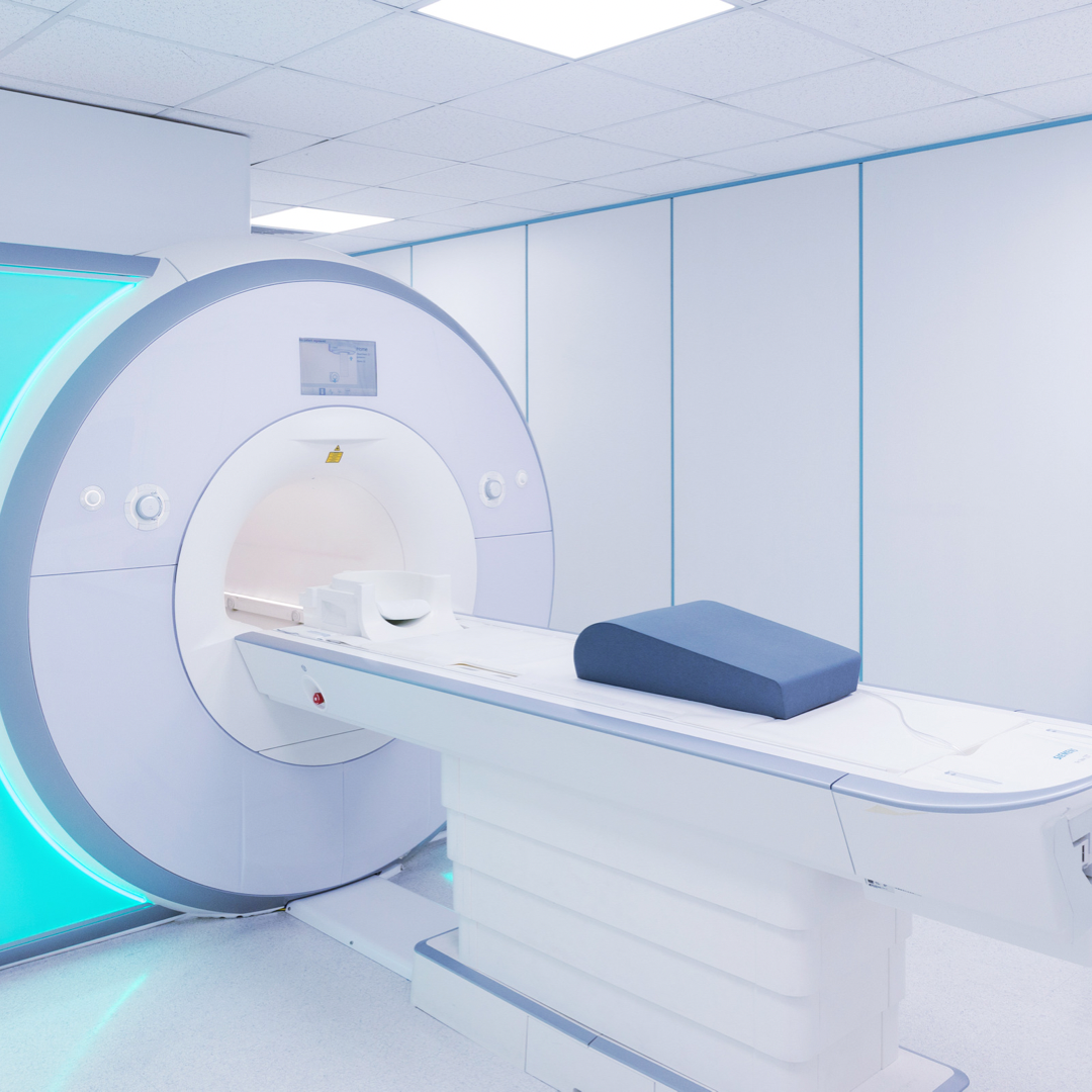
To utilize some of the imaging tests that we have, such as MRIs, I generally will wait until x-rays are done, flexion-extension, as I described. Anti-inflammatory medications, Naprosyn twice a day for three to four weeks, not intermittently, is a good one to try, and a complete. cervical spine physical therapy program to strengthen the neck muscles. If, in fact, this doesn’t work over three or four weeks, and one is convinced that pressure on a nerve root, for instance, the biceps muscle is weak or the wrist extensors are weak and you definitely have a radiculopathy, I will put somebody on a course of steroids. Dexamethazone, four milligrams, four times a day for two days, three a day for two days, two a day and one a day for two days for a total of eight days, this basically will reduce the swelling of the nerve root stabilizing the cervical spine with cervical traction to sleep in at night is something I’ve done on a repetitive basis.
For cervical traction, one puts a little pad underneath the chin and the occiput, and you hang five pounds over the end of the bed and sleep in it. What this does is it puts slight tension on the cervical spine. If you take a chain and you lay it on a table, you can move it back and forth. If you put a little bit of tension on the chain, it doesn’t move, so effectively with a minimal amount of traction to sleep in overnight, as tolerated, the motion in the cervical spine is diminished, allowing the steroids to work. This has been a very fruitful, positive direction for me. Is it a permanent? Well, that’s to be determined, but if you can relieve the pain, that’s fine. If all these fail, then an MRI would be indicated, but to get an MRI and shoot from the hip is confusing to most untrained doctors who have a predatory nature to make money for their institution.
When and only when would a surgery such as anterior decompression or spinal fusion, known as an ACDF be required for a cervical disc?

Specifically, for surgery, anterior cervical discectomy and fusion is done when all this has failed, and the radiculopathy is unchanged. A 95% success rate for radiculopathy is expected. I was trained to take bone from the hip. That’s basically negatively stated now by most people, because no one knows how to take bone from the hip. You relegate your first year resident and they chop up the muscles and it hurts a lot. And that’s not true, that doesn’t work. Technique is imperative! The industry of spine has come up with allografts. In other words, bone from someone else is put in the cervical disc. The idea is to spread the disc apart, slightly, take the disc material out, put a block in there, a bone block to spread it apart. And if you distract the foramina in the back where the bone spur is, it doesn’t move anymore, it doesn’t traumatize the nerve root. That in itself is a classical operation, and a safer one.
This has been enhanced by the industry of spine to put cages and bone morphogenic protein and even plates at this point. I did lots of surgeries in the seventies, eighties and nineties, with just bone from the hip, and it works. To do more is really overkill and it just creates an increase in costs. So the idea as discussed by Robbie Robinson with me, when I was a young man, at breakfast one morning as described, one puts the bone graft in after taking the disc out, evacuate it completely. He never decompressed the disc in the back, never went into the back. This is for pure radiculopathy, and spreading it apart stabilizes it, decompresses the nerve root, and if one’s, carpentry is correct, that bone graft will be lodged in place within the semi hemispheric curvatures of the cervical disc above and below. And it locks in place. The plates are done, I think it’s overkill. It creates more of a problem. There are complications with this surgery so the shorter you make it, and the less complicated you make it, the greater the chance of doing well.
What is the recovery time from an ACDF?
The recovery time from an ACDF, when I did no plates and just bone graft from the hip, I’d see them in two weeks. I’d take the sutures out of the neck. Generally, it was a subcuticular suture with paper sutures so there’s really little scar. I’d probably put them in a collar for two to four weeks until their neck pain resolved. Then I would leave them out of a collar. I get an x-ray at six weeks and one at three months, the incorporation of the bone graft is very clear. About the sixth or eighth week I would have them walking and doing regular, relatively benign exercises. And if the neck muscles are weak, physical therapy can strengthen the neck muscles. This was not a diagnosis of weak muscles, this was a diagnosis of radiculopathy. Once that’s resolved, if therapy is necessary for the specific muscle group of the upper extremities, then that would be the thing to focus on.
Conclusion
To recap all this and cervical disc is a clinical diagnosis of radiculopathy, generally caused by a bone spur, not by a soft disc as it is in the lumbar spine. Over-reading an MRI with a “bulging cervical disc” and having three herniated discs is really not truthful. That’s a normal finding. There are papers out to show that cervical MRIs can be done in normal people, and 35-40% of them have something you could operate on. That’s a real boon for someone trying to make a living, but it’s not good for the patient. So once this diagnosis is made, conservative treatment of steroids, anti-inflammatory medications, cervical traction, I would hesitate to put people in a cervical collar as that will weaken the muscles. If it’s tolerated without a collar, I would not use one, but the cervical traction is important. I had great results with that.
Once all this is exhausted, then I would also add a flexion-extension film to make sure there’s no excessive motion at that one level. And if there’s none, then it’s already fused. Your chances of working and doing it conservatively are excellent. Moving on, to get an MRI at this point in time to see what we’re dealing with, an MRI will show a bulging disc, but does not necessarily show an osteophyte. An oblique x-ray of the cervical spine will show a foramen to be obliterated with a bone spur on the oblique side left and right, and I used those for years to make that diagnosis. So an MRI will help confirm what’s going on. And in rare instances, a cervical radiculopathy with objective clinical findings, not just complaints of arm pain, which is subjective, you’ll end up having a surgical procedure completed which will resolve the problem. One has to have very clear objective findings in order to go ahead and proceed with any surgery at this time.


